The pnCCD Color X-Ray Camera® (CXC)
The Color X-Ray Camera (CXC) is PNDetectors’ high resolution spectroscopic X-ray imaging system based on our energy dispersive pnCCD detector and, optionally, polycapillary optics. The CXC is a stand-alone system with a complete electronic and data acquisition system, a full software package enabling camera operation and monitoring, data-acquisition and live visualization of the X-ray spectrum and spatially resolved elemental images. The camera can be operated in ambient conditions, as well as configured such that it can be integrated into a vacuum system, e.g. at a synchrotron beamline.
The CXC combines the following key performance parameters in a single instrument:
-
4D imaging: simultaneous time-, energy- and spatial resolution (xny)
-
Ultra-fast readout of up to 1000 fps
-
Wide energy range (200 eV - 30 keV)
-
Superior energy resolution for all X-ray events (145 eV for 6 keV)
-
High count rate of up to 106 stored counts/s
-
Outstanding quantum efficiency (80% for 500 eV, 90% for 10 keV, 30% for 20 keV)
-
Several external trigger and sync signal
 is PNDetector’s high resolution spectroscopic X-ray imaging system based on an energy dispersive pnCCD detector")
The Color X-Ray Camera (CXC) is PNDetector’s high resolution spectroscopic X-ray imaging system based on an energy dispersive pnCCD detector

pnCCD with its readout ASICs

CXC System

pnCCD® Properties
Highlights
Working Principle

Overview
FF-microXRF
X-Ray Micro-Tomography
Polychromatic SAXS
Simultaneous X-ray Fluorescence and Diffraction
CXC System Overview
Camera model options
-
Stand-alone model with Beryllium window for operation under ambient pressure
-
Vacuum solution
-
Optional: polycapillary optics which can be used for either configuration
All CXC camera models provide the following system components:
pnCCD® Sensor
-
Active sensor area of 12.7 mm × 12.7 mm
-
Pixel: 264 × 264 (69,696) full frame area, plus frame store area
-
Dimension of camera head: 120 mm × 212 mm × 80 mm (W × H × D)
-
Various operation modes
-
For details see (pnCCD Properties)
Complete electronic system and data acquisition system
-
Mountable in 19 inch rack
-
High speed parallel data acquisition
-
Selectable amplifier bandwidth and analog gain for high dynamic range
Full software package for
-
Camera control
-
Live visualization
-
Offline data analysis
High precision X-ray optics (optional)
-
Exchangeable poly-capillary optics
-
Focusing, defocusing and 1:1 with capillary diameters from 15μm to 30μm

pnCCD CXC Camera - front and back view

Polycapillary optics
pnCCD Properties
| Physical pixel size | 48 µm × 48 µm × 450 µm |
|---|---|
| Number of physical pixels | 264 × 264 (69,696) full frame area, plus frame store area |
| Active area | 12.7 mm × 12.7 mm (161 mm2) full frame area |
| Frame rate | up to 1,000 Hz (264 × 264 pixel) |
| Pixel readout rate | up to 70 MegaPixels/s |
| Windowing mode | 24 × 264 pixels (smallest window) |
| Externally triggerable | yes |
| Readout noise (rms) | ENC typ. 3e- / pixel at 100Hz, 200Hz ENC typ. 4e- / pixel at 400Hz ENC typ. 8e- / pixel at 1000Hz |
| Energy resolution | typ. 145 eV for Mn-Kα |
| Sub-pixel spatial resolution | Δx < 15 µm (rms) for 1 keV X-rays |
| Charge handling capacity | up to 400,000 signal electrons per pixel |
| Radiation hardness | up to 1014 photons/cm2 at 10 keV |
Performance Highlights
The CXC system is equipped with a 264 × 264 pixel pnCDD with a physical pixel size of 48 µm × 48 µm and an imaging area of 161 mm2. The on-chip electronics in combination with a fully column-parallel readout enables low noise operation at about 3 e-/pixel (ENC), and frame rates of up to 1000 Hz. Due to the advanced detector design, the internal potential distribution of the pnCCD can be adjusted by the operation voltages depending on the requirements of the individual experiments. Thus, excellent energy resolution (145 eV for Mn-Kα) can be achieved, as well as high charge handling capacity (up to 400000 e- per pixel), and anti-blooming modes for handling extremely high amounts of signal charges.
Spectroscopic performance
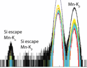
Excellent elemental information between 200 eV and 30 keV with an energy resolution of 145 eV (FWHM) at 5.9 keV (Mn-Kα) and 83 eV at 277 eV (C-K).

Spectrum of a radioactive 55Fe source
Ultra-fast readout and excellent signal-to-noise ratio
Standard full frame rate of 100, 200, 400, 1,000 fps, up to 8,000 fps using windowing and binning. Excellent signal-to-noise ratio even at high readout speeds with typical 3e-at 100 fps up to 8e- at 1,000 fps.
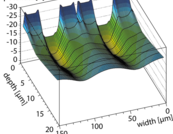
High dynamic range
The CXC is capable to provide a HCHC mode (High Charge Handling Capacity, up to 400,000 e- per pixel), as well as an anti-blooming mode for handling extremely high amounts of signal charges. The impact of the HCHC mode is depicted below:
Inimitable single photon sensitivity (left image) and high intensity imaging (right image) with 400,000e- per pixel (corresponding to 10³ photons with an energy of 1 keV) at the same time.

Single photon (left) and high intensity images (right)
Subpixel Resolution
Depending on the experimental requirements, some of our customers are interested in a spatial resolution beyond the CXC pixel size of 48 µm. This subpixel resolution can be realized with two approaches, which can also be combined.
-
The first approach, the so-called "optical subpixel resolution", reaches this goal by equipping the CXC with magnifying policapillary optics or by using pinhole optics. A spatial resolution of approx. 8 µm was demonstrated with a CXC equipped with a magnifying 6:1 polycapilary optics [B. De Samber et al. (2019)]. Compared to a polycapillary optics, a pinhole optics provides the capability for "zooming", i.E. to vary the magnification factor according to the experimental needs, however at the expense of lower detector count rates. For a detailed evaluation of both methods of optical subpixel resolution with the CXC please refer to B. De Samber et al. (2019).
-
The second approach is the so-called "computational subpixel resolution". This method is based on dedicated data post-processing. A spatial resolution better than 3 µm at 1.3keV was demonstrated with this method by S. Ihle et al. (2017), please refer to this publication for a detailed discussion. As the native CXC frms6 raw data format provides the full raw data information, it enables the user to apply such a method e.g. as an optional data post-processing "on demand" on any native CXC raw data set which meets the requirements regarding the count rate and photon statistics.
pnCCD® Heritage
The pnCCDs have an outstanding heritage in diverse fields of science. For example, two projects based on the pnCCDs which provided excellent data and a multitude of highly rated publications in journals with high impact factor like nature, are the EPIC camera and the CAMP instrument.
-
The pnCCDs are the core element of the EPIC camera on board of the European X-ray observatory XMM-Newton [Soltau (1996), Strüder (2001)], which was launched in 1999 and is still operative. The excellent quality of the EPIC data led to several thousand publications.
-
A ground based application of the pnCCD is the CAMP instrument which was installed at the LCLS (Linac Coherent Light Source) at SLAC for several years (see e.g. [Strüder (2010), Chapman (2011), Seibert (2011), Loh (2012), Rudek (2012), Johansson (2012)]) and was afterwards installed at FLASH at DESY.

The pnCCD® Principle
The pnCCDs are back illuminated, three-phase CCDs on a fully depleted silicon substrate. Their operation is based on the principle of sideward depletion and on transfer registers formed by pn-junctions. The outstanding characteristics include a homogeneous and thin photon entrance window leading to high quantum efficiency values between 100 eV and 20 keV and an excellent radiation hardness as well as high charge handling capacity.
Due to its excellent spatially and spectroscopically resolving capabilities, it is possible to use the CXC for a broad variety of applications, for example:
-
XRF (X-ray flouresence spectroscopy)
-
Full-field XRF, FF-µXRF with polycapillary optics
-
Simultaneous XRD (X-ray diffraction) and XRF
-
TXRF (total reflection XRF)
-
Elemental mapping
-
XANES (X-ray absorption near-edge spectroscopy) / NEXAFS (near edge X-ray absorption fine structure)
-
GEXAF (grazing exit X-ray absorption flouresence)
-
CT / µCT (X-ray computed tomography)
-
Polychromatic SAXS (small-angle X-ray scattering)
-
Fluctuation X-ray scattering (FXS)
-
Transmission X-ray imaging
-
Laue X-ray diffraction
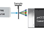
With the pnCCD® based CXC, high-performance energy-resolved X-ray imaging is available for a wide range of experimental and laboratory conditions. A multitude of experiments can be performed with a single camera for a broad range of samples. An interesting example for the versatility of the CXC is its application in the field of Full-field µXRF (FF-μXRF). For this method, a broad beam X-ray source illuminates the entire sample, and the CXC preserves the spatial coherence of the image using polycapillary optics. The X-ray data is assembled into an X-ray spectrum image, where a full X-ray spectrum is saved at each individual pixel. From this, the elemental peaks can be identified, and false-color elemental images can be formed. Images are made without scanning the sample, which also removes all difficulties that come with the alignment of a focused, primary X-ray beam. As a result, FF-μXRF is flexible enough to image uneven surfaces, dangerous or toxic materials and samples in an aqueous solution that are difficult to analyze with μXRF. Moreover, FF-μXRF allows the detection of single, locally concentrated elements in one view. In order to increase the spatial resolution beyond the pixel size of the pnCCD, the CXC can be equipped with magnifying polycapillary optics.
The Color X-ray Camera (CXC) is PNDetector’s high resolution spectroscopic X-ray imaging system based on an energy dispersive pnCCD detector

FF-microXRF

X-Ray Micro-Tomography

Polychromatic SAXS

Simultaneous X-ray Fluorescence and Diffraction
Full-Field Micro X-ray Fluorescence (FF-µXRF)
The CXC system in combination with a poly-capillary optics provides a spatially resolved elemental image of the sample. This can for example be used to image biological samples. A water flea (daphnia magna) was imaged at the BAM line at the BESSY synchrotron. The internal organs and overall structure of the insect are clearly visible, as the CXC system allows wet, small, unpolished specimens to be analyzed with trace level sensitivity and high position resolution.
Combining the CXC with a magnifying polycapillary enhances the spatial resolution beyond the physical pixel size of the pnCCD®. It was demonstrated, that by applying a 6:1 polycapillary optics, a resolution of 8µm was achieved. An alternative solution for enhanced spatial resolution is the application of a pinhole optics in front of the CXC [see B. De Samber et al. (2019) for a detailed discussion of both options].
FF-microXRF

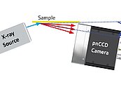
With an X-ray source at a high incident angle, the CXC system enables imaging of samples with large topographic features with minimal shadowing. Thus, in places like the eyes on the statue shown below, the traces left behind by ancient paint can be identified. The iron based pigments show up as traces in this area of the statue, along with a lead based pigment in the middle. A flexible imaging system based on the CXC is capable of measuring many different types of samples, from flat, polished specimens, to rough samples with little to no sample preparation [I. Reiche (2013)].

Therefore the FF-µXRF technique satisfies the requirement of artists and archeologists for imaging and analysis of cultural heritage objects. It allows analysts to quickly identify critical areas and hidden structures, with the advantage that the imaging technology is extremely similar to other cameras commonly used for optical, UV-VIS and NIR imaging. The final result is always an image that contains spatial and spectroscopic information.
X-Ray Micro-Tomography
In case where self-absorption of the sample is low, e.g. low-absorbing biological samples, Full Field –XRF (FF-XRF) projections can be recorded for various orientations of the sample. This allows to reconstruct a 3D image from the two dimensional FF-XRF images, the so-called µCT technique.
As a matter of course, due to the capability of the CXC to record energy dispersive images with the full spectroscopic information stored for every pixel of the images, the generation of various element-specific 3D sinograms from one µCT data set is possible. Combining the CXC with a magnifying polycapillary enhances the spatial resolution beyond the physical pixel size of the pnCCD [De Samber (2019)].

Polychromatic SAXS
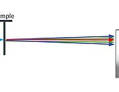
Most Small Angle X-ray Scattering (SAXS) systems require an X-ray source that is monochromatic. Since monochromation is inherently inefficient, these systems require expensive monochromators and high flux X-ray systems. In contrast, with the CXC, no monochromation is required. The resulting X-ray data can be energy filtered or processed to create diffraction patterns or reciprocal space images. As shown in the SAXS images from a sample of silver behenate (AgC22H43O2), monochromatic SAXS will result in the typical ring pattern (right image). Although the rings are larger in the polychromatic case (left image), the CXC enables energy dependent analysis of the diffraction rings, resulting in a faster measurement with more data and high angular resolution.

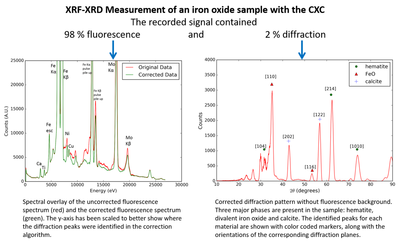
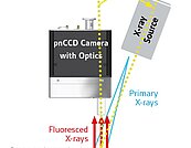
Simultaneous X-ray Fluorescence and Diffraction
Due to the fact that the CXC records both the position and energy of each incoming X-ray photon, high speed, high resolution X-ray diffraction experiments are possible in transmission as well as in traditional forward scattering modes. At every single point in the image, a full diffraction pattern and fluorescence spectrum can be derived. This is done simultaneously, without monochromatization, stage, scanning or beam focusing, and the measurements take less than one minute to acquire. Owing to the detectors’ capability to record spatially and spectroscopically resolved images, the simultaneous XRD-XRF signal can be separated, resulting in diffraction patterns without fluorescence background and fluorescence spectra without diffraction peaks.